Texture Methods for Histology Image Analysis
Project Details
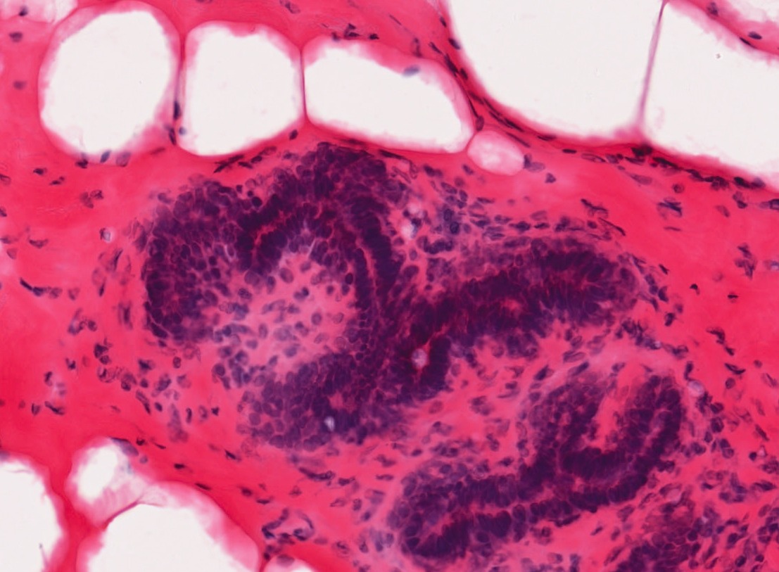
Background: Recent advances in digitizing whole microscope slides have enabled the creation of large databases of images. Examining each image by hand to determine the composition of different tissue types and inspect for anomalies is both inefficient and subjective, even for a trained pathologist. With the development of image analysis techniques, the work can be automated and quantitative measures standardized. This project explored texture methods for classifying tissue types in histology images. Such images are taken by a microscope of tissue, such as that obtained by biopsy. Prior to imaging, sections of tissue are stained to increase the contrast of structures of interest. Hematoxylin and eosin (H&E) is one such stain which turns nuclei blue and cytoplasm pink. The magnification is high enough that cell structures such as nuclei and membranes are visible and different tissue types can be identified.
Solution: Through the use of color and texture analysis methods, the classification of tissue types was automated. The data set used in this work consisted of breast tissue that had been stained with H&E. Three different tissue types of interest were labeled and used to develop a classifier that could later be used for segmentation. This project explored color and texture methods for characterizing the appearance of tissue. The standard approach to texture classification involves convolving the image with a set of filters and clustering the responses into textons. This technique was compared with a variation involving quantile functions and methods to measure local image curvature.
Additional Applications: The texture methods used in this project can be used for classifying or segmenting any type of image. In a later project they were used to initialize a tissue segmentation routine.